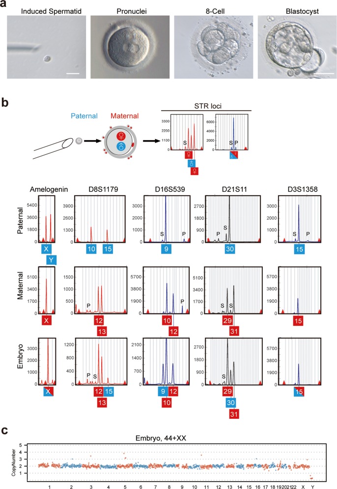
Fig. 5. Functional analysis of spermatids derived from in vitro testicular organogenesis.
a A MII oocyte derived through IVM was injected with an induced spermatid, followed by development to pronuclear stage with two pronuclei, 8-cell and blastocyst stages. Scale bars = 10 µm (left); 100 µm (right). b STR and sex loci of embryo, and fetal skin (paternal) that contributed the round spermatid and granulosa cells (maternal) of oocyte. S: Stutter peak; P: Pull down peak. c CNV analysis of the embryo (b) by genomic DNA sequencing.

