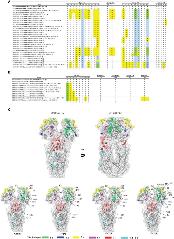
Figure 4.
Alignment of the antigenic epitopes of VP4 of the Lebanese RVAs with those of Rotarix® and RotaTeq®. Representative Lebanese strains corresponding to those in the VP4 tree are shown. (A) Alignment of the antigenic epitopes of the VP8 of the vaccines and representative RVAs circulating in Lebanon. (B) Alignment of the antigenic epitopes of the VP5 of the vaccines and representative RVAs circulating in Lebanon. Residues that are marked by yellow-color are residues that are different from Rotarix® and RotaTeq®, in green-color are residues that are different from the respective genotypic strain in RotaTeq®, and blue-color are the residues that are different from Rotarix®. Amino acid residues known to mediate escape from neutralization with mAbs (2) are indicated by an asterisk (*). (C) Surface representation cartoon of the VP4 protein (PDB 3GZT). The right image is rotated 90° compared to the left image. Antigenic epitopes are colored in green (8–1), blue (8–2), yellow (8–3), pink (8–4), red (5–1), and cyan (5-2 to 5-5). Substitutions relevant to parent vaccine strains are shown in spheres.

