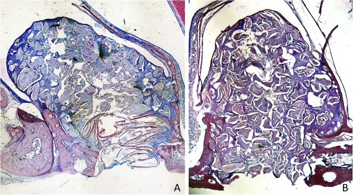
Fig. 3.
Photomicrographs of decalcified sections illustrating the healing after 1 week. a Treated site. Bone strips occupying the antrostomy and the subjacent area (close-to-window region). b Untreated site. Note the new bone-forming from the sinus bone walls. Scarlet-acid fuchsine and toluidine blue stain. Images grabbed at × 20 magnification

