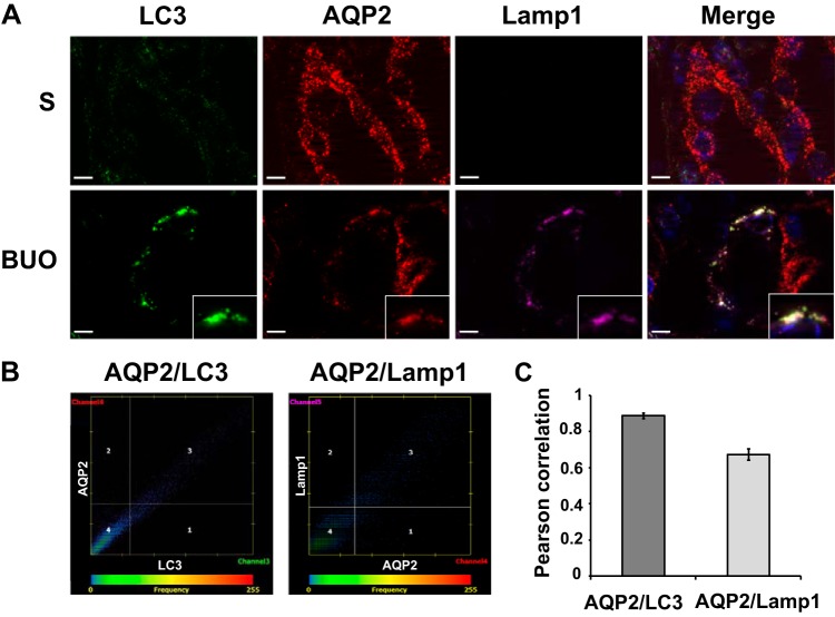
Fig. 3.
Colocalization of aquaporin 2 (AQP2) with autophagy markers in inner medullary collecting duct (IMCD) cells of 4-h bilateral ureteral obstruction (BUO) and sham (S) rats. A: inner medulla sections of sham (n = 3) and 4-h BUO (n = 3) were triple labeled against AQP2 (red), lysosomal associated membrane protein 1 (Lamp1; pink), and microtubule-associated protein 1A/1B-light chain 3 (LC3; green). Insets: magnified views of the areas where significant colocalizations were observed as a group of puncta that were not seen in control sections. Scale bars = 4 µm. B: representative scatterplots demonstrating the high degree of colocalization between AQP2 and LC3 (Pearson correlation coefficient, r2 = 0.90, P < 0.05) or Lamp1 (r2 = 0.67, P < 0.05) in IMCD cells of a rat with 4-h BUO. C: bar graph summarizing Pearson correlation coefficients of AQP2 and LC3 or Lamp1 from 30 colocalized puncta. Two independent experiments were performed.

