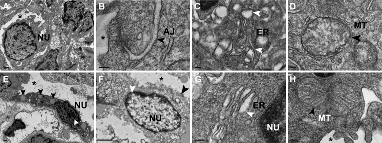
Fig. 6.
Structural abnormalities in inner medullary collecting duct (IMCD) cells of 10- and 24-h bilateral ureteral obstruction (BUO) rats. A: electron micrograph showing the condensed cytoplasm, severely deformed IMCD cells, and collapsed tubular lumens (*) in 10-h BUO rats (n = 3). Scale bar = 1 µm. B–D: IMCD cells of 10-h BUO rats (n = 3) contained disrupted adherens junctions (AJ; black arrowhead; B), enlargement of the endoplasmic reticulum (ER) lumen (white arrowheads; C), and slightly enlarged mitochondria (MT) with loss of membranes (black arrowhead; D). Scale bars = 100 nm. E–H: as for 24-h BUO rats (n = 3), IMCD cells contained many lipid droplets (black arrowheads) in a shrunken cytoplasm and pyknotic nuclei (NU; white arrowhead). Scale bars = 1 µm. IMCD cells demonstrated rupture of nuclei (white arrowhead; E), rupture of epithelium tubular lumens (black arrowheads; F), enlargement of the ER lumen and loss of the ER membrane (white arrowhead; G), and loss of the mitochondrial membrane and cristae (black arrowhead; H). Scale bars = 100 nm. Two independent experiments were performed.

