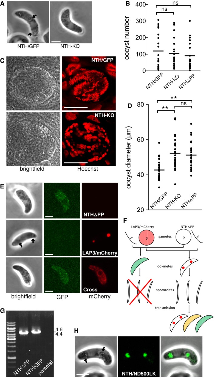
Brightfield images of a NTH‐KO and NTH/GFP ookinete, showing normal ookinete morphology, but lack of pigment clusters associated with crystalloids (arrows) in the NTH null mutant. Scale bar = 5 μm.
Aligned dot plot of oocyst numbers in Anopheles stephensi infected with parasite lines NTH/GFP, NTH‐KO, and NTHΔPP. Horizontal lines mark mean values (n = 20); ns, not significantly different (Mann–Whitney).
Brightfield and fluorescence images of oocysts of parasite lines NTH/GFP and NTH‐KO at 15 days post‐infection, showing lack of sporulation in the NTH null mutant. Hoechst DNA staining (artificially depicted red) labels parasite nuclei. The large nuclei of the epithelial midgut cells also show. Scale bar = 20 μm.
Aligned dot plot of oocyst diameters in A. stephensi at 15 days post‐infection with parasite lines NTH/GFP, NTH‐KO, and NTHΔPP. Horizontal lines mark mean values (n = 20). ** mark statistically significant differences (P < 0.0001, Mann–Whitney); ns, not significant.
Images of ookinetes of parasite lines NTHΔPP showing dispersed NTHΔPP::GFP fluorescence; LAP3/mCherry showing focal LAP3::mCherry fluorescence in crystalloids; and cross (carrying modified alleles encoding NTHΔPP::GFP and LAP3::mCherry), showing dispersed LAP3::mCherry fluorescence. Arrow marks pigment cluster associated with crystalloids. Scale bar = 5 μm.
Schematic diagram of a genetic cross between parasite lines NTHΔPP and LAP3/mCherry and the resultant ookinete population after mosquito transmission and drug selection (oocysts, liver, and blood‐stage parasites not shown). Colours represent NTH::GFP (green), LAP3::mCherry (red), and their co‐localisation (orange). Spots in ookinetes depict crystalloids. The red cross indicates that sporozoite formation is blocked. Self‐fertilisation events are omitted.
PCR diagnostic for the presence of modified nth alleles in blood‐stage infections obtained after sporozoite transmission of a NTHΔPP x LAP3/mCherry genetic cross (lane NTHΔPP), amplifying an ˜ 4.4 kb fragment. NTH/GFP parasites are used as a PCR control, amplifying ˜ 4.6 kb. Parental parasites provide a negative control.
Image of a NTH/ND500LK ookinete, showing fluorescent spots co‐localising with pigment clusters (arrows) indicative of normal crystalloid formation. Scale bar = 5 μm.

