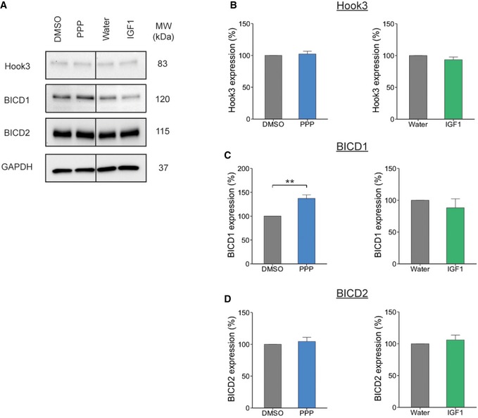
Figure 5. IGF1R inhibition alters the expression level of BICD1.

- Western blot of PMN treated with 1 μM PPP or 50 ng/ml IGF1 for 60 min. Cells were lysed and the extracts assessed for expression levels of dynein adaptor proteins, all proteins were assessed from the same blot.
- Hook3 expression did not change after treatment with PPP or IGF1 compared to controls (DMSO, 100%; PPP, 102.16 ± 4.3%, P = 0.67, Student's t‐test, N = 3 independent experiments; water, 100%; IGF1, 93.48 ± 4.2%, P = 0.26, N = 3 independent experiments; data shown are mean ± SEM).
- However, BICD1 expression was significantly increased in PMN treated with PPP compared to control (DMSO, 100%; PPP, 137.1 ± 7.3%, **P = 0.002, Student's t‐test, N = 7 independent experiments). In contrast, IGF1R stimulation by IGF1 had no effect on BICD1 expression (water, 100%; IGF1, 88.1 ± 14.0%, P = 0.49, Student's t‐test, N = 3 independent experiments; data shown are mean ± SEM).
- BICD2 expression did not change after treatment with PPP or IGF1 compared to controls (DMSO, 100%; PPP, 104.44 ± 6.5%, P = 0.51, N = 3 independent experiments: water, 100%; IGF1, 106 ± 7.5%, P = 0.51, Student's t‐test, N = 3 independent experiments; data shown are mean ± SEM).
Source data are available online for this figure.
