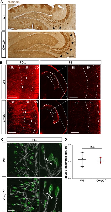
Figure EV3. Related to Fig 3. Axon pruning in crmp2 −/− mice is altered in Sema3F‐related but not Sema3A‐related regions.

- Calbindin immunostaining of sagittal brain sections shows defects in pruning similar to those seen in coronal sections of WT and crmp2 −/− mice in Fig 3. Arrowheads indicate the infrapyramidal bundle. Scale bars: 100 μm.
- Hippocamposeptal axons are pruned in both WT and crmp2 −/− mice (n = 3). At P0–1, retrogradely DiI‐labeled CA1 pyramidal neurons showed strong signal in both WTs and crmp2 −/− indicating the presence of CA1 projections into the medial septum. (Arrowheads indicate retrogradely labeled cell bodies.) Conversely, at P8, no retrogradely labeled CA1 cell bodies were detected in CA1 granular layer in either WT or crmp2 −/− mice. SR, SP, and SO indicate stratum radiatum, stratum pyramidale, and stratum oriens, respectively. Scale bars: 100 μm.
- Maximum intensity projections of P11 triangularis sterni muscle NMJs of WT and crmp2 −/− mice (n = 3). Axons were stained with βIII‐tubulin antibody (white) and postsynaptic acetylcholine receptors with α‐bungarotoxin (green). Boxed areas are shown on the right at higher magnification with examples of doubly innervated NMJs where two axons are competing for synaptic innervation (arrowheads). Scale bars: 50 μm.
- Quantification of the number of doubly innervated NMJs revealed no significant difference (WT 4.5 ± 1.8%, cmrp2 −/− 4.2 ± 0.7%, P > 0.99, n = 3 pups/genotype). Mean ± SD, Mann–Whitney test.
