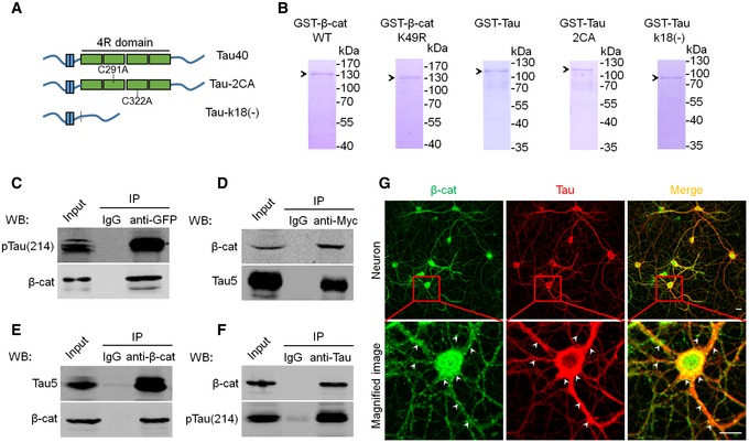
Figure EV3. Affinity‐purified Tau and β‐catenin proteins and the association of Tau with β‐catenin in vitro and in vivo .

-
AThe schematic shows full‐length Tau (Tau40), and the acetyltransferase activity domain double‐mutated Tau (cystine‐291/322 to alanine, Tau‐2CA) or the activity domain‐deleted Tau (Tau‐k18(−)).
-
BThe representative Coomassie blue staining pattern of the affinity‐purified β‐cat WT, β‐cat K49R, Tau40, Tau‐2CA, and Tau‐k18(−) from Escherichia coli using Ni‐NTA resin.
-
C, DAssociation of Tau with β‐cat in vitro. HEK293 cells with stable expression of Tau40 were transfected with eGFP‐β‐cat (C); or naïve HEK293 cells were transiently transfected with myc‐Tau40 (D). After 48 h, the cell extracts were immunoprecipitated using anti‐GFP or anti‐myc followed by Western blotting using Tau5, pS214, and β‐cat antibodies, respectively.
-
E, FAssociation of Tau with β‐cat in vivo. The mouse hippocampal extracts (3‐month‐old) were immunoprecipitated using anti‐β‐cat (E) or Tau5 (F) followed by Western blotting using Tau5 or pS214 and β‐cat antibodies, respectively.
-
GCo‐localization of Tau and β‐cat in primary hippocampal neurons infected with AAV‐Tau detected by co‐immunofluorescence labeling (scale bars, 10 μm); white arrowheads indicate co‐localization of Tau and β‐cat.
Source data are available online for this figure.
