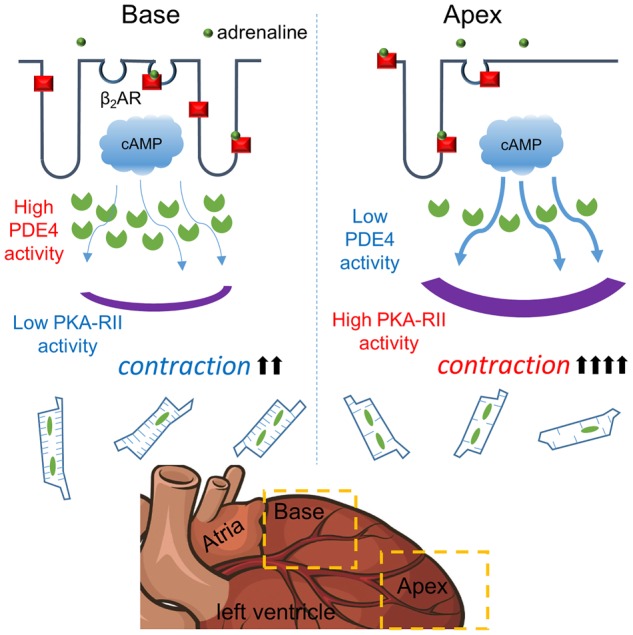
Figure 2. Diagram demonstrating the variability of cardiomyocyte membrane organisation depending on localisation within the LV (apical or basal myocardial regions).

Within the basal region, LV cardiomyocytes present a highly organised structure, which is associated with restricted cAMP diffusion, while within the apex cardiomyocytes cAMP produced upon β2AR activation is more far-reaching. In apical cardiomyocytes β2AR can enhance cellular contractility but in basal cells cAMP is restricted by greater compartmentation. (Reproduced from Wright et al. [41]).
