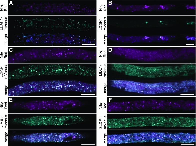
Figure 5.
Subcellular localization of selected candidate LD proteins in N. tabacum pollen tubes. A to F, Candidate proteins fused to mVenus at their C termini were transiently expressed in N. tabacum pollen tubes (cyan channel). LDs were stained with Nile red (magenta channel). In the merge channel, colocalization appears white. Note, in B, that expression of LDDH1-mVenus led to clustering of LDs. Bars = 10 μm.

