Figure 5.
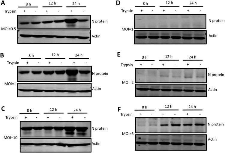
LLC-PK cells were more susceptible to PDCoV infection than ST cells. LLC-PK cells were infected with PDCoV at an MOI of (A) 0.5, (B) 1, or (C) 10, and ST cells were similarly infected at an MOI of (D) 1, (E) 2, or (F) 5. Both infected cell types were cultured in the presence or absence of 5 μg/ml trypsin and then cells were washed and lysed for western blot at 8, 12 and 24 hpi. PDCoV N proteins were analyzed with a specific antibody against N protein, and actin was used as a loading control. Experiments were repeated at least three times.

