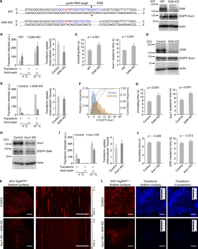
Figure S4.
Effects on clathrin-mediated endocytosis of knockout or knockdown of GAK and knockdown of Aux1. (A) CRISPR/Cas9 gene editing strategy used to knock out GAK in cells gene edited for EGFP-Aux1+/+ and CTLA-TagRFP+/+. The double strand break (red triangles) induced by Cas9 resulted in elimination of four nucleotides (dotted lines). Loss of GAK expression was confirmed by Western blot with antibodies against GAK, Aux1/GAK, and actin (right). (B) Effect of GAK knockout on receptor-mediated uptake of transferrin (n = 3 experiments, mean ± SD). (C) Uncoating time and Aux1 residence time, in cells lacking GAK. Data from bottom surfaces of double-edited EGFP-Aux1+/+ and CLTA-TagRFP+/+ cells with GAK (n = 5 cells) or lacking GAK by knockout (n = 7 cells) imaged at 1-s intervals for 200 s by TIRF microscopy (mean ± SD, P values by two-tailed t test). (D) Western blot analysis of EGFP-Aux1+/+ and CLTA-TagRFP+/+ cells treated with lentivirus containing control shRNA (Control) or shRNA specific for GAK (GAK-KD), showing specific reduction of GAK expression 5 d after transduction. (E) Effect of GAK knockdown on receptor-mediated uptake of transferrin (n = 2 experiments, mean ± SD). (F) Influence of GAK depletion on Aux1 recruitment. Data from bottom surfaces of double gene-edited EGFP-Aux1+/+ and CLTA-TagRFP+/+ cells with GAK (1,058 traces, eight cells) or depleted of GAK by knockdown (1,380 traces, nine cells) imaged at 1-s intervals for 200 s by TIRF microscopy. The number of recruited EGFP-Aux1 molecules is significantly increased (Cohen’s d = 0.68). (G) Influence of GAK depletion on uncoating time (left) and Aux1 residence time (right). Data from bottom surfaces of double-edited EGFP-Aux1+/+ and CLTA-TagRFP+/+ cells with GAK (n = 5 cells) or depleted of GAK by knockdown (n = 5 cells) imaged at 1-s intervals for 200 s by TIRF microscopy (mean ± SD, P values by two-tailed t test). (H) Western blot analysis of parental SUM159 cells incubated with lentivirus containing control shRNA or shRNA specific for Aux1 (Aux1-KD) showing specific reduction of Aux1 expression 5 d after transduction. (I) Effect of Aux1 knockdown in gene-edited EGFP-GAK+/+ and CTLA-TagRFP+/+ cells on receptor-mediated uptake of transferrin (n = 2 experiments, mean ± SD). (J) Influence of Aux1 depletion on uncoating time (left) and GAK residence time (right). Data from bottom surfaces of double-edited EGFP-GAK+/+ and CLTA-TagRFP+/+ cells with Aux1 (n = 5 cells) or depleted of Aux1 by knockdown (n = 5 cells) imaged at 1-s intervals for 200 s by TIRF microscopy (mean ± SD, P values by two-tailed t test). (K) Bottom surfaces of AP2-TagRFP+/+ cells treated with lentivirus containing control shRNA or a mixture of shRNA targeting Aux1 and GAK (Aux1-KD + GAK-KD) imaged at 2-s intervals for 300 s by spinning-disk confocal microscopy. The representative images are from a single time point; the corresponding kymograph shows the entire time series. Scale bars, 10 µm. (L) AP2-TagRFP+/+ cells with or without double Aux1 + GAK knockdown incubated with 10 µg/ml Alexa Fluor 647–conjugated transferrin for 10 min at 37°C and then imaged in 3D using spinning-disk confocal microscopy (30 imaging planes spaced at 0.35 µm). Images from the bottom surface of control cells show diffraction-limited AP2-TagRFP spots associated with endocytic coated pits and coated vesicles; in cells depleted of Aux1 and GAK, the punctate distribution is replaced by characteristic larger patches. The images also show the extent of surface binding (bottom surface) and internalization (maximum z-projection of the 30 stacks) of transferrin in the control cells and its absence in the cells impaired in endocytosis due to the Aux1 and GAK depletion. Scale bars, 10 µm.

