Figure 9.
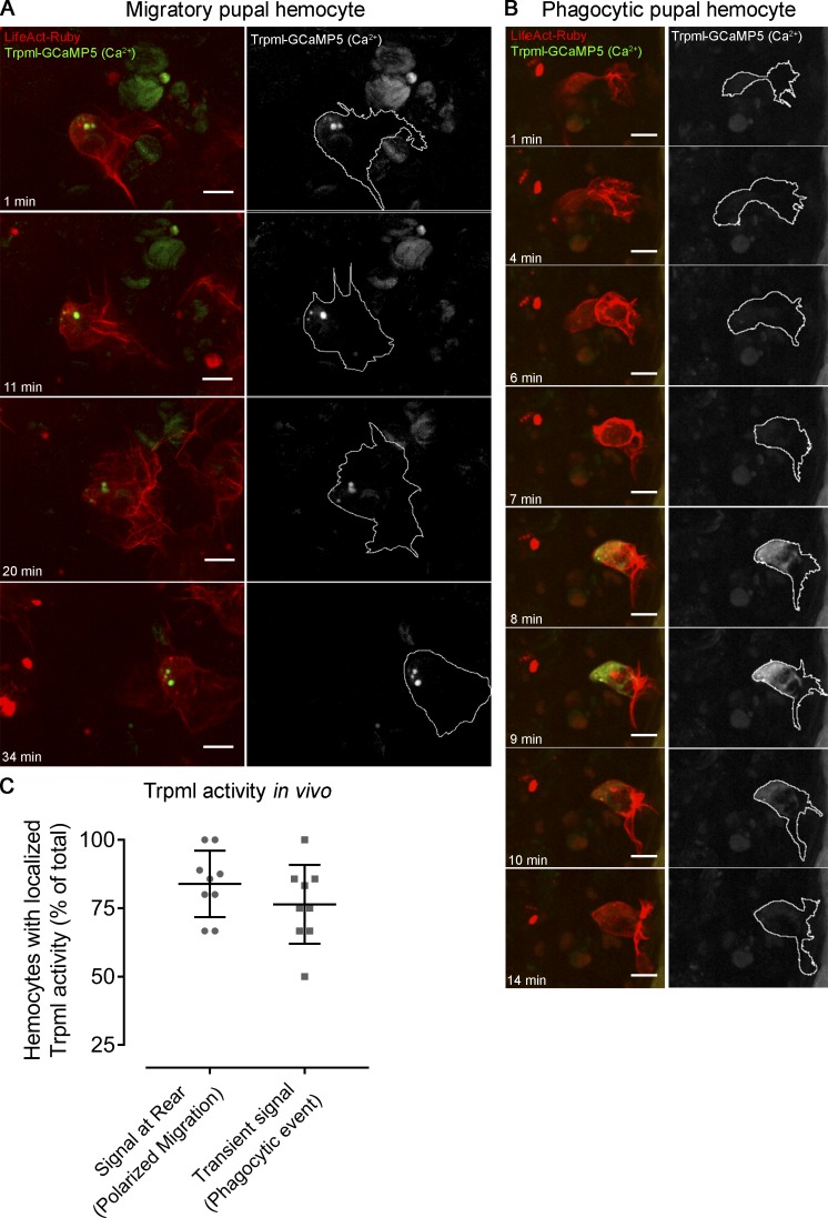
Trpml activity in vivo. Representative images of mitotic clones labeling pupal hemocytes expressing LifeAct-Ruby and calcium sensor trpml-GCaMP5, revealing the activity of the channel in vivo. (A) Migrating hemocyte containing signal at the rear, which perdures over time, suggesting activity of Trpml from the lysosomes. (B) Hemocyte generating a protrusion where transient spikes of activity arise, which probably corresponds to a phagocytic event. Scale bar in A and B: 10 µm. Full videos are included in Video 9. (C) Proportion of migrating cells with signal at the rear and transient spikes of activity upon engulfment. Values are presented as scatter dot plot indicating mean ± SD (n ≥ 4 independent experiments, n ≥ 2 pupae/experiment).

