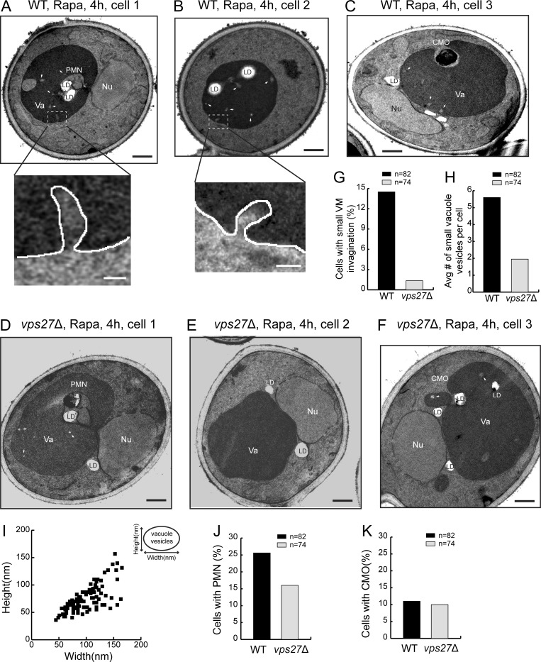
Figure 4.
The ESCRT deletion abolished one form of microautophagy. (A–C) Representative TEM images showing three types of microautophagy, including PMN, CMO, and small VM invagination in WT cells after rapamycin (Rapa) treatment. Insets are zoomed-in images showing the small tubular invagination of VM. (D and E) Representative TEM images showing that after Rapa treatment, PMN and CMO still happened in vps27Δ cells, while the small VM invagination was nearly abolished. (G) Frequency of observing small VM invagination in WT and vps27Δ cells after Rapa treatment. (H) Number of small vacuole vesicles per cell in WT and vps27Δ cells after Rapa treatment. (I) Size distribution of small vacuole vesicles. (J) Frequency of observing PMN in WT and vps27Δ cells after Rapa treatment. (K) Frequency of observing CMO in WT and vps27Δ cells after Rapa treatment. LD, lipid droplet; Nu, nucleus; Va, vacuole. White arrowheads highlight small vacuole vesicles. Black scale bar, 0.5 µm; white scale bar, 50 nm.

