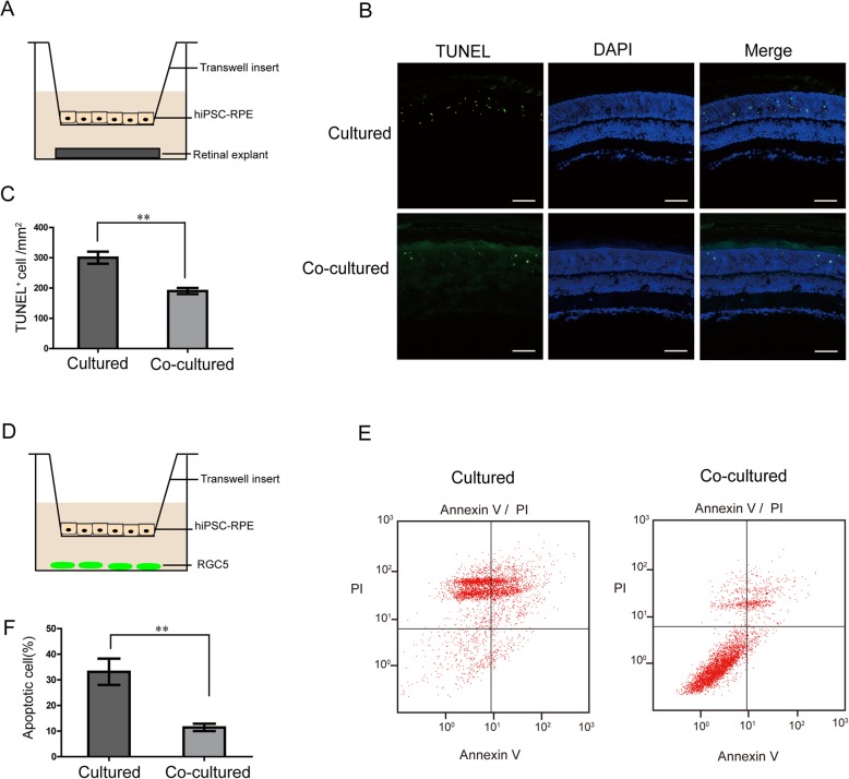
Fig. 2.
Protective effect of co-culturing of retinal explants and RGC5 with hiPSC-RPE in vitro. a Model of co-culture system: hiPSC-RPE cells co-cultured with retinal explants separated by transwell insert. b TUNEL/DAPI co-staining of the retinal explant after co-culturing with and without hiPSC-RPE cells after 2 days. c Bar chart representing the positive TUNEL staining of cells per square millimeter compared between retinal explants with or without co-culturing. d Illustration of the transwell co-culture system: RGC5 co-cultured with hiPSC-RPE cells. e Flow cytometry analysis of apoptosis in RGC5 co-cultured with and without hiPSC-RPE. f Bar chart shows the rate of apoptosis in percentage compared between RGC5 with and without co-culturing. Scale bar 50 μm (b). Data are presented as mean ± SEM. p values were determined by unpaired two-tailed Student’s t test (c, f), n = 4 for each group, *p < 0.05, **p < 0.01

