Fig. 5.
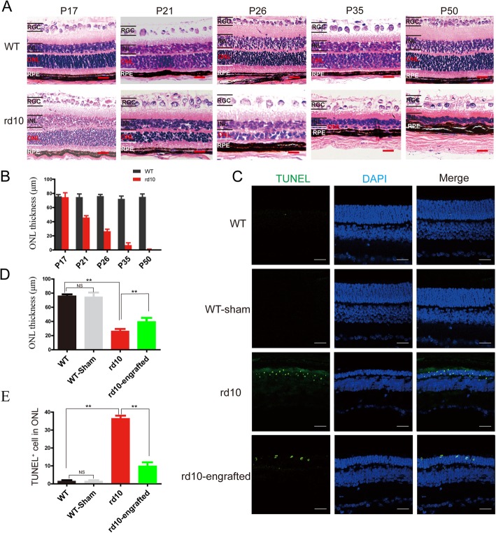
hiPSC-RPE cells preserved of photoreceptor in rd10 mice. a Histology of rd10 and WT mouse retina over time. b Quantification of the retinal outer nuclear layer thickness in rd10 and WT. c Retinal outer nuclear layer thickness and apoptotic cells in the retina of hiPSC-RPE-transplanted rd10 detected by DAPI and TUNEL staining, retinas from non-transplantation rd10 mice, and WT at the same age are used as a comparison. d Quantification of TUNEL-positive cell density. e Analysis of retinal outer nuclear layer thickness of after hiPSC-RPE cell transplantation. Scale bar 20 μm (a), 50 μm (c). Data are presented as mean ± SEM. p values were determined by one-way ANOVA followed by Tukey multiple comparison tests (d, e), n = 5 for each group, *p < 0.05, **p < 0.01

