
Figure 2.
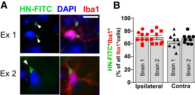
Intravenously administered HN is found in microglia in the mouse brain. A, HN-FITC (1 μg/g in PBS) was injected via the femoral vein 24 h after ICH onset. After another 24 h, confocal microscopy was used to detect HN-FITC (green, white arrowheads) in the ICH-affected brain (ipsilateral) and their colocalization with Iba1+ cells (red). Representative examples of two randomly selected HN-FITC-positive cells are shown. Nuclei were stained with DAPI (blue). Scale bar, 10 μm. B, Quantitative bar graph (mean ± SEM) showing the percentage of HN-FITC/Iba1 double-positive cells among all the Iba1+ cells, as assessed in nine or 10 randomly selected perihematoma (ipsilateral) and homologous contralateral locations (Contra) in two independently analyzed mouse brains.