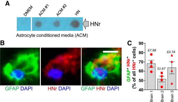
Figure 4.
Astrocytes in culture secrete HN, and HN is present in astrocytes in the brain. A, Rat ACM was concentrated using Vivaspin and spotted onto nitrocellulose membranes. HN (2 μl from 10 mm HN stock) and DMEM were used as positive and negative controls, respectively. Image represents two different ACM samples from each culture that were spotted. Extracellularly released HN was detected using anti-HNr (a rat homolog of HN) antibody. B, A strong HNr immunofluorescence signal (red) was observed in GFAP+ (green) rat astrocytes. Confocal image. Scale bar, 6.25 μm. C, Quantitative bar graph (mean ± SEM) showing the percentage of HNr/GFAP double-positive cells among all the HNr+ cells, as assessed in four randomly selected locations in three independently analyzed mouse brains.

