Figure 5.
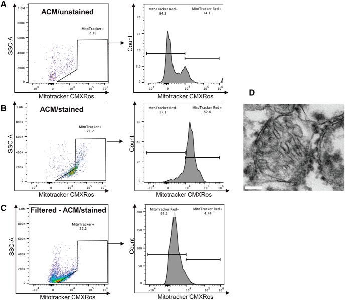
Mt are released by astrocytes in vitro. Rat cortical astrocytes were incubated for 30 min with Mitotracker/Red/CMXRos to label Mt. After extensive washing, astrocytes were incubated with fresh culture medium for 24 h to accumulate Mt in the media. ACM was collected and briefly spun down to remove cellular debris and assessed for the presence of Mitotracker-positive Mt using FACS analysis. A, Negative control, ACM from astrocytes that were not stained with Mitotracker/Red/CMXRos. B, ACM collected from astrocytes stained with Mitotracker/Red/CMXRos. C, ACM collected as in B, but filtered through a 0.22 μm pore-size column to remove Mt (Filtered-ACM). FACS analysis data for the above samples. D, An example of Mt found in ACM as detected using TEM. Scale bar, 100 nm.

