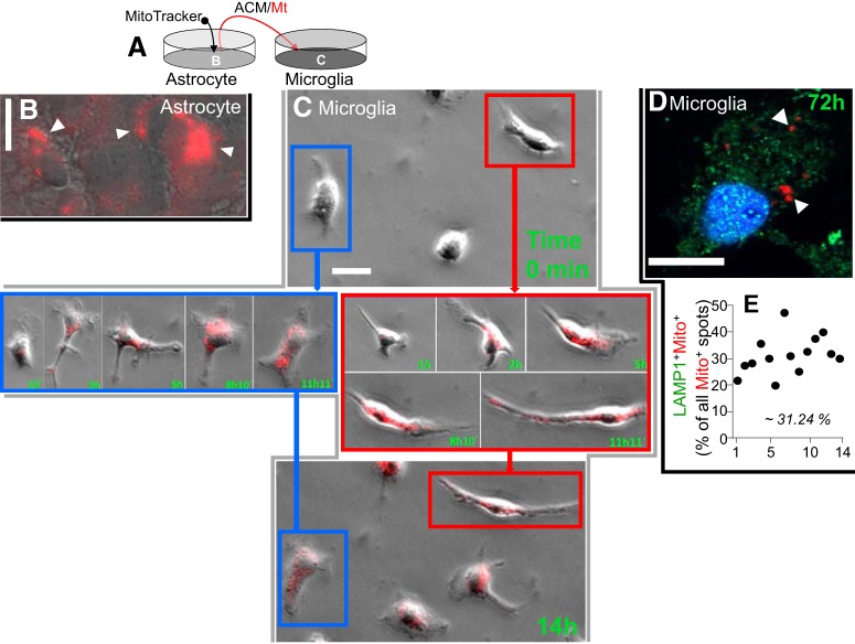
Figure 6.
Astrocyte-secreted Mt incorporate into microglia and elevate intracellular HN levels. A, Astrocytes in culture were treated with MitoTracker/CMX/Ros to red-fluorescently label Mt. After extensive washing to remove MitoTracker and additional 24 h incubation to accumulate Mt in the media, the media containing red-labeled astrocytic Mt were transferred to rat primary microglia in culture. B, Representative confocal image of rat astrocytes in culture treated with MitoTracker/CMX/Ros to label Mt (red puncta, white arrowheads). Scale bar, 20 μm. C, Microglia taking up astrocytic Mt. Time-lapse images were collected to document Mt (red) transfer into microglia over a 14 h period. Images of two representative microglial cells (color-coded as blue and red) before Mt transfer (0 min, top) and at the indicated times (15 min to 11 h, middle; and at 14 h, bottom). Scale bar, 20 μm. At the end of the recording, all the microglial cells showed the presence of MitoTracker (Figure 6-1). D, Confocal image of microglial cells containing astrocytic Mt (MitoTracker red+ puncta). These astrocyte-derived Mt (red, white arrowheads) are retained in microglia for at least 72 h after internalization. Most of the Mt (Mito+ red puncta) did not colocalize with the LAMP-1 (green puncta), suggesting that Mt are not merely undergoing lysosomal degradation. Scale bar, 10 μm. E, Quantitative data showing the percent of LAMP-1/MitoTracker double-positive puncta among all the MitoTracker red+ puncta, as determined for 14 randomly selected microglial cells.

