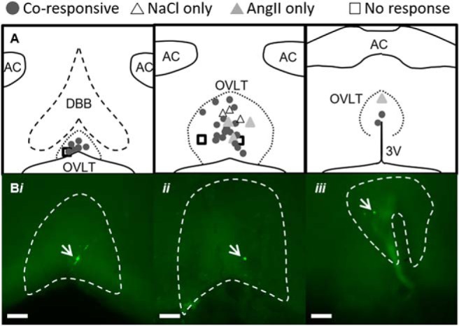
Figure 3.

Neuroanatomical map of OVLT neuron responses to hypertonic NaCl and AngII in vitro. A, Schematic illustration of three rostrocaudal levels and anatomical location of OVLT neurons recorded in vitro and presented in Figure 2. B, Examples of neurobiotin-labeled and coresponsive neurons (green, indicated with white arrows) at three different anatomical locations within OVLT: (Bi) rostromedial, (Bii) lateral margin, (Biii) dorsal cap. Scale bars, 100 μm (10× images).
