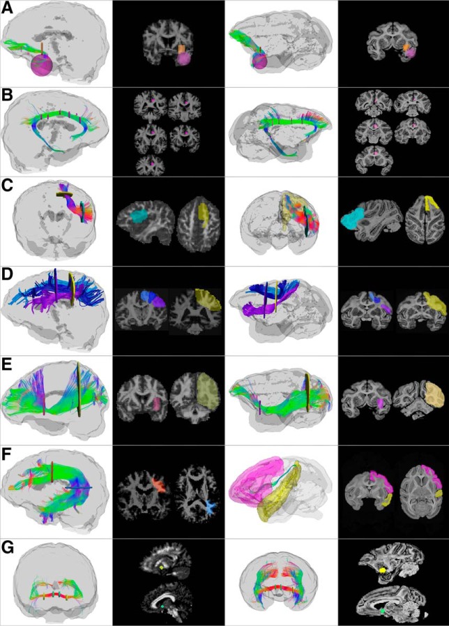
Figure 2.
Regions of interest used to dissect individual tracts in the human (two left columns) and monkey (two right columns) brain. For each example, 3D reconstructions and 2D sections are shown. In addition to the regions depicted here, exclusion regions were used in the midsagittal plane, brainstem, subcortical nuclei, and internal capsule to exclude commissural and projection tracts and remove individual spurious streamlines. A, Uncinate fasciculus (lateral view). Inclusion regions of interest are placed in the anterior temporal lobe (pink) and external/extreme capsules (orange). B, Cingulum (medial view). A single inclusion region (pink) on multiple coronal slices along the cingulate gyrus is used to ensure that all the short projections of the dorsal cingulum are included. C, Frontal aslant tract (anterior view). An inclusion region (light blue) is placed in the white matter medial to the inferior frontal gyrus in the sagittal plane. In humans, a second inclusion region (yellow) is placed in the white matter inferior to the superior frontal gyrus in the axial plane, whereas in monkeys, an atlas-defined region of the superior frontal gyrus is used as the second region to include all streamlines projecting to the medial frontal regions. Exclusion regions were then placed in the frontal pole. D, SLF (lateral view). Posteriorly, one inclusion region (yellow) is placed in the parietal lobe in line with the superior aspect of the central sulcus, whereas anteriorly three separate inclusion regions are used for each of the three branches: SLF I (light blue), II (dark blue), and III (purple), all in a coronal plane passing through the precentral gyrus. Exclusion regions are used in the temporal and occipital lobe in both humans and monkeys. E, Inferior fronto-occipital fasciculus (lateral view). One inclusion region is used in the external/extreme capsules (pink) and one in the anterior border of the occipital lobe (yellow); both are in the coronal plane. F, Arcuate fasciculus, long segment. In the human, one inclusion region (orange) is placed in the coronal plane just anterior to the central sulcus, and one inclusion region in the axial plane inferior to the temporoparietal junction (blue). In the monkey, to be as inclusive as possible, atlas-defined regions of the frontal lobe (pink mask) and superior temporal gyrus (yellow mask) were also used as inclusion regions of interest. In addition to the inclusion regions pictured here, exclusion regions were placed in the external/extreme capsules and the white matter of the superior temporal gyrus to remove the middle longitudinal fasciculus, and in the white matter medial to the supramarginal gyrus to remove SLF fibers. G, Anterior commissure. Two inclusion regions were used to capture the compact bundle of the anterior commissure as it crosses the midline. Each region has two slices in the sagittal plane on either side of the midline, one more medial (green), one placed more laterally (yellow). Exclusion regions were used to remove spurious streamlines forming part of the fornix, anterior thalamic projections, and other projections from the brainstem.

