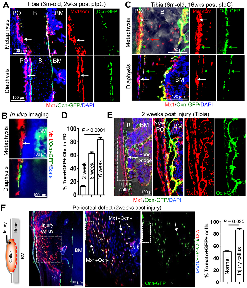
Figure 2. Mx1+ progenitors in the periosteum supply the majority of osteoblasts in cortical bone injury.
A-C, Using 3-month old (A & B, 2 weeks after 5 doses of pIpC induction) and 6-month old (C, 16 weeks after pIpC induction) Mx1/Tomato/Ocn-GFP mice (IR and WT-BMT at 4 weeks of age), periosteal Mx1+ progenitor cells (white arrows) and endosteal osteoblasts (GFP+), new osteoblasts (Tomato+GFP+, green arrows) and osteocytes (red arrows) from Mx1+ progenitor cells (C) in the tibia were analyzed by anti-GFP staining (A & C) and intravital imaging (B) (n=3–5 per group). PO, periosteum; B, bone; BM, bone marrow. D. Percentage of Mx1+Ocn+ (Tomato+GFP+) periosteal osteoblasts and osteocytes in the metaphysis and diaphysis of the tibia 2 (A), 8 (data not shown), and 16 (C) weeks after pIpC induction (n=5). E & F. Representative tibia drill-hole defect (E) or periosteal defect (F) on cortical bone surface of Mx1/Tomato/Ocn-GFP mice. Contribution of Mx1+ periosteal progenitors (Tomato+) to the most of the new Mx1+Ocn+ osteoblasts (Tomato+GFP+) in the bone bridge (E) and to the majority of callus-forming osteoblasts (Mx1+Ocn+; white arrow) in the cortical defect (F) were assessed. White boxes in E and F represent the panels to the right. Graph shows percentage of Mx1+Ocn+ osteoblasts from uninjured periosteum (normal) and injured bone callus (Injury callus) (n=5).

