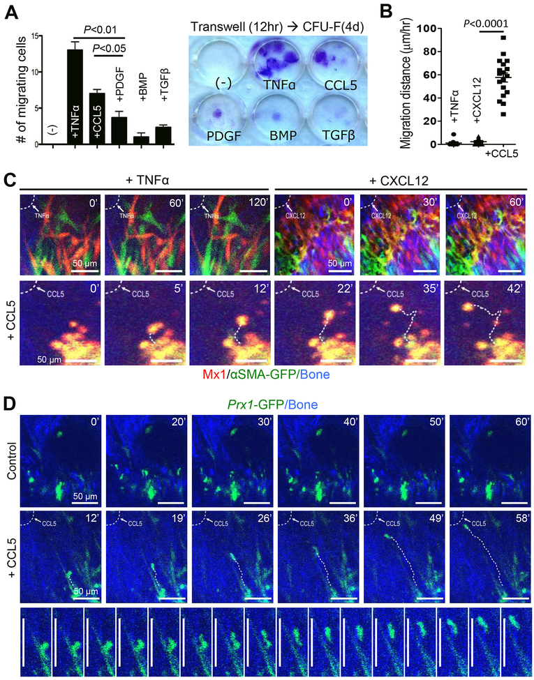
Figure 5. Migration of P-SSC is induced by CCL5 in vitro and in vivo.
A. The effects of TNF-α (10 ng/mL), CCL5 (20 ng/mL), PDGF-β (20 ng/mL), BMP (20 ng/mL), and TGF-β (10 ng/mL) on the migration of sorted Mx1+Ocn− periosteal cells (CD105+CD140a+Mx1+Ocn-GFP− cells from Figure S2E; 5,000/well) were examined using a transwell assay (8-μm pore) for 12 hours (left), followed by colony formation (>50 cells) for 4 days (right) (≥ 3 replicates). *P<0.05; **P<0.01. B. The average in vivo migration distance of individual Mx1+αSMA+ P-SSCs induced by TNFα, CXCL12, or CCL5 (all, 2 L at 10 ng/μL of Matrigel) was measured in Mx1/Tomato/αSMA-GFP mice from Figure 1A. C. In vivo real-time imaging shows that CCL5, but not TNFα or CXCL12, at the injury site (dotted line) induced the migration of Mx1+αSMA+ P-SSCs (Tomato+GFP+). The numbers indicate the time of imaging. Representative of 3–5 mice per group. D. CCL5 induces in vivo real-time migration of Prx1+ P-SSCs (GFP+) in Prx1CreERGFP mice treated with CCL5 (2 μL at 10 ng/μL of Matrigel) or Matrigel alone (2 μL, Control). Bottom images represent sequential images of Prx1+ P-SSC migration over 16 minutes (1 image per minute, representative of 5 mice). Scale bar is 50 μm.

