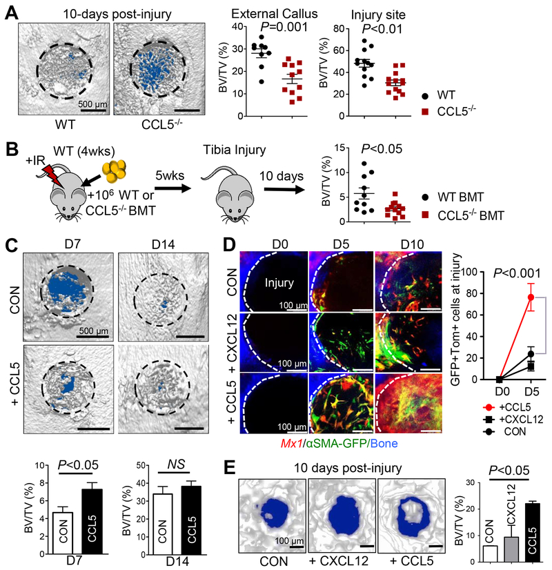
Figure 6. CCL5 is necessary for Mx1+αSMA+ P-SSC recruitment and activation leading to accelerated bone healing.
A. Representative μCT images of proximal tibial injuries (left) of age and sex-matched WT (n=12) or 2- to 4-month old CCL5−/− mice 10 days post-injury (n=13). BV/TV of external callus (left) and injury sites (right) was assessed. B. WT-BL6 mice (n=10 per group) were irradiated (9.5 Gy) and transplanted with 106 WT (WT-BMT) or Ccl5−/− BM mononuclear cells (Ccl5−/− BMT). Five weeks later, BV/TV of injury sites was assessed at 10 days after tibial drill-hole injuries. C. Representative μCT images (top) and BV/TV quantification (bottom) of tibia injuries (WT-BL6 mice) treated locally with CCL5 (+CCL5; 10 μL at 1 ng/μL Matrigel) or under control conditions (10 μL Matrigel, CON) on Days 0, 2, and 4 post-injury (n=5–7). D. Sequential in vivo imaging of 3- to 4-month old Mx1/Tomato/αSMA-GFP mice from Fig. 1 treated with supplemental CCL5, CXCL12 (2 μL at 10 ng/μL of Matrigel), or Matrigel alone (2 μL, CON) at the indicated times after calvarial injury. E. Representative μCT images (left) and BV/TV (right) analysis of calvarial injuries (from 6D) treated with CCL5, or CXCL12, 10 days post-injury (left).

