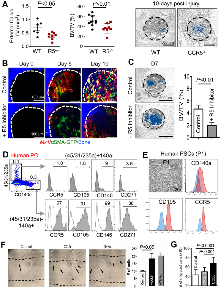
Figure 7. CCR5-CCL5 influences periosteal cell migration and bone healing.
A. The external callus volume (left), BV/TV (right), and μCT images of proximal tibial bone injuries in WT or Ccr5−/− mice at 10 days post-injury were used to assess bone healing. B-C. Sequential in vivo imaging of Mx1/Tomato/αSMA-GFP mouse (3-month-old) calvarial injuries treated with a CCR5 inhibitor (+R5 Inhibitor; 10 μL at 2.5 μg/μL Maraviroc in Matrigel) or gel control (10 μL of Matrigel) for 10 days post-injury (B). Representative μCT images on Day 7 post-injury (C, left) and BV/TV analysis (C, right graph) were used for the quantification of bone healing. D. Primary cells isolated from human periosteal tissue (Human PO) were analyzed for expression of CCR5 and indicated SSC markers. Dotted line, isotype controls. E. Single-cell driven colonies from human periosteal cells were cultured for 7–10 days (P1), then analyzed for expression of CCR5 and SSC markers (CD105 and CD140a). Blue line, isotype controls. F. A scratch assay was used to determine the effects of 10 ng/mL of CCL5 or TNFα on human periosteal cell migration (10,000 cells/well). G. A transwell migration assay (8-μm pore) was used to determine the effects of CCL5 (5 ng/mL) or Cxcl12 (5 ng/mL) on human periosteal migration (P1, 15,000/well) migration for 24 hours.

