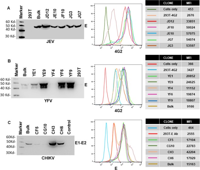
Figure 4.
Generation of stable cell lines secreting JEV, YFV and CHIKV VLPs. 293 T cells were transduced with (A) JEV (B) YFV or (C) CHKV structural protein containing lentivirus particles. Transduced cells were bulk selected with blasticidin followed by limiting dilution single cell cloning. For each virus, several single cell clones were characterized for Envelope protein staining on the cell surface via flow cytometry and VLP secretion into the culture supernatants via western blotting. Mean Fluorescence intensity (MFI) of Envelope protein staining for the different single cell clones for JEV, YFV and CHIKV is shown on the right. Gel images were analyzed using GENETOOLS gel analysis Software version 4.03 (f) (Syngene, https://www.syngene.com/software/genetools-automatic-image-analysis/). Flow cytometry histograms were created using the FloJo Software version 10.6.0 (Tree Star, https://www.flowjo.com/solutions/flowjo/downloads).

