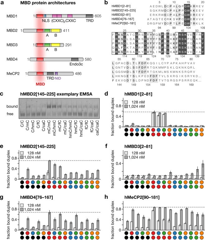
Figure 2.
MBDs and DNA recognition. (a) Protein architecture of five MBD family proteins. (b) Amino acid sequence of human MBD domains used in this study. Identical residues in dark gray and residues with similar properties in light gray; residues in vicinity to the DNA duplex are framed. (c) Representative EMSA of hMBD2[145–225]. (d–h) Bar diagrams of the fraction of MBD-bound DNA duplex observed in EMSA for MBDs and nucleobase combinations as indicated. Data are means ± SEM from three independent experiments (see SI for full data).

