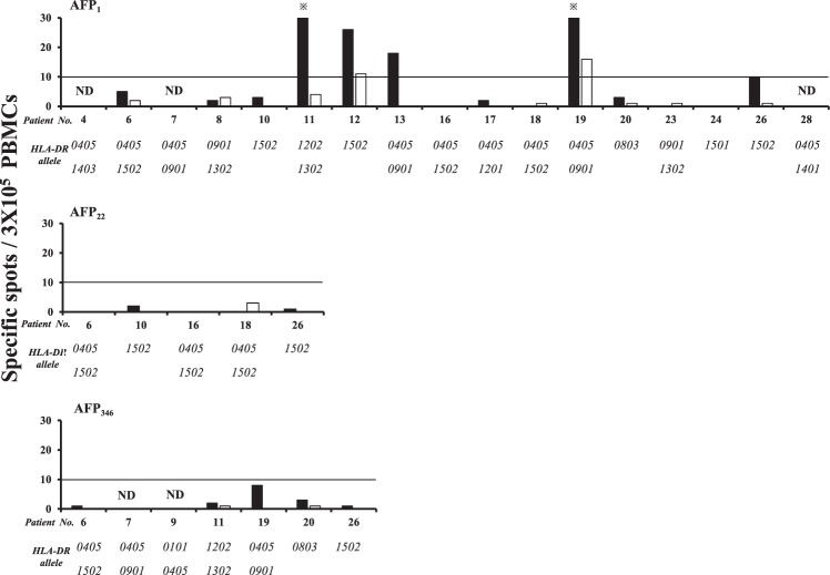
Figure 2.
Responses of CD8+ and CD4+ cell-depleted PBMCs to AFP-derived epitopes. The numbers of spots on IFN-γ ELISPOT and the HLA-DR alleles of each patient are shown. Black and white bars denote the results for CD8+ and CD4+ cell-depleted PBMCs, respectively. The positive rate of CD8+ cell-depleted PBMCs against AFP1 was 35.7% (5/14), while that of CD4+ cell-depleted PBMCs was 14.3% (2/14). ND denotes “not determined”. *indicates >30 spots.

