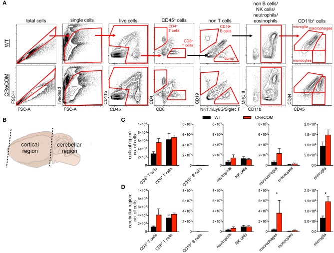
Figure 3.
Evidence of diffuse leukocyte infiltration in CReCOM brains. Brains of 11-12-month-old female WT (n = 4) and CReCOM (n = 4) mice were split into two portions and analyzed by flow cytometry. Data are representative of three independent experiments. (A) Whole brain gating strategy. (B) Regional breakdown for subsequent analysis. (C) Analysis of cortical region infiltration of T-cells (CD4+CD45hi and CD8+CD45hi), B-cells (CD19+CD45hi), neutrophils (CD11b+Ly6G+CD45hi), NK cells (CD11b+NK1.1+CD45hi), macrophages (CD11b+CD64+CD45hi), monocytes (CD11b+CD64−CD45hi), as well as microglia (CD11b+CD64+CD45lo). (D) Analysis of aforementioned populations in cerebellar region (*p < 0.05).

