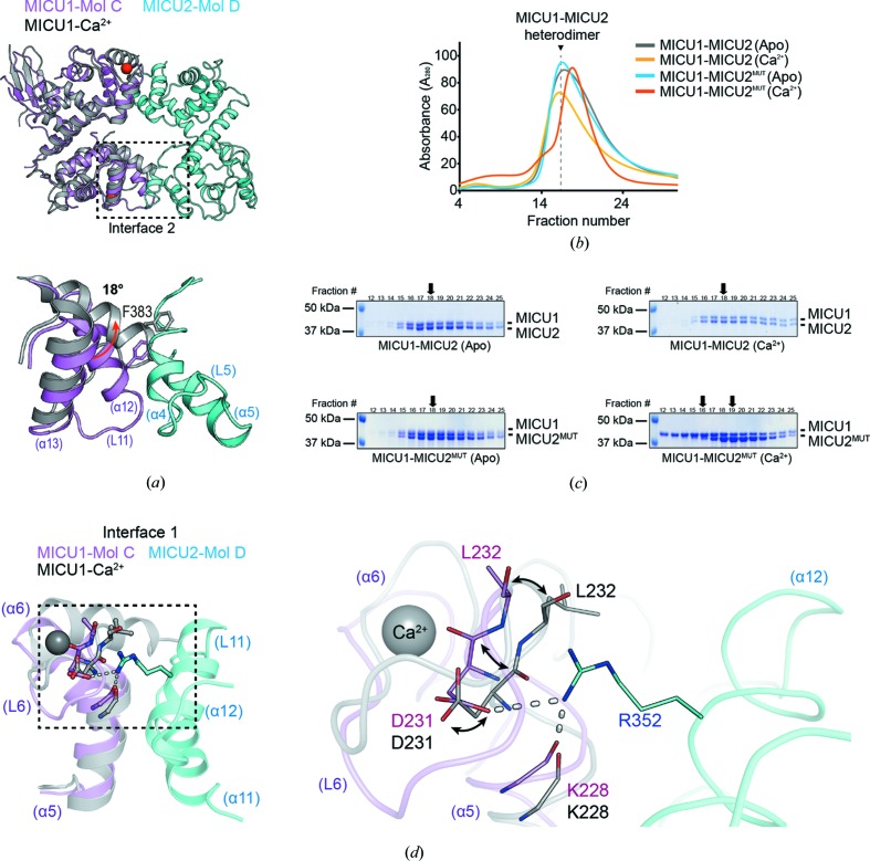
Figure 4.
Structural and biochemical analysis for comparison with Ca2+-bound MICU1. (a) Cartoon representations of the superimposed apo heterodimer and one molecule of Ca2+-bound MICU1 (gray) (PDB ID 4nsd; Wang et al., 2014 ▸), and a detailed view of interface 2 of the superimposed structure. The 18° tilt of α12 helix of MICU1 EF-hand 3 is indicated by a red arrow. The heterodimer is colored violet (MICU1) and cyan (MICU2). (b) SEC profile of the MICU1–MICU2 or MICU1–MICU2MUT heterodimer, and (c) its SDS–PAGE results in the absence (marked by Apo) or presence of Ca2+ (marked by Ca2+). The black arrows indicate the peak fractions of each SDS gel. The two black arrows in the MICU1–MICU2MUT(Ca2+) heterodimer indicate the peak fractions of MICU1 and MICU2 (left and right), respectively. (d) A cartoon representation of the superimposed interface 1 of the apo MICU1–MICU2 heterodimer with one molecule of Ca2+-bound MICU1 (gray) (PDB ID 4nsd) based on MICU1 in the heterodimer. The conformational changes of MICU1 EF-hand 1 including the Asp231 and Leu232 residue are indicated by a stick representation and black arrows.

