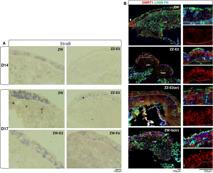
Fig. 4.
The expression pattern of mitotic-meiotic switch markers is affected in the cortical germ cells of embryos subject to oestrogen levels alteration. (A) RNA in situ analysis of STRA8 expression on sections from the left gonad of D14 (HH40) and D17 (HH43) embryos. ZW, female wild-type control; ZZ-E2, ZZ treated with β-oestradiol at D7-7.5 (HH31); ZW-E2, ZW treated with β-oestradiol at D7-7.5 (HH31); ZW-Fa, ZW treated with fadrozole at D7-7.5 (HH31). STRA8 expression is severely compromised in the gonadal cortical germ cells from ZZ embryos exposed to β-oestradiol. (Ba-Bl) Immunofluorescence detection of DMRT1 (red), LHX9 (green) and Fibronectin 1 (FN; blue) on gonadal cryostat sections from the following D17 (HH43) embryos: ZW, female wild-type control (Ba-Bc); ZZ-E2, ZZ treated with β-oestradiol at D7-7.5 (HH31) (Bd-Bf); ZZ-E2(sr), ZZ treated with β-oestradiol at D4 (HH23) (partially sex reversed) (Bg-Bi); ZW-Fa(sr), ZW treated with fadrozole at D4 (HH23) (partially sex reversed) (Bj-Bl). Right-hand panels show high-magnification images of the boxed areas on the left; (Bb,Be,Bh,Bk) LHX9, FN and DMRT1; (Bc,Bf,Bi,Bl) DMRT1 only. All samples were cut sagittally (left gonad shown) apart from ZZ-E2, which was cut transversally (left and right gonads shown). White dotted lines mark the cortex-medulla border. In the left wild-type ovary, LHX9 marks cortical somatic cells, FN highlights the cortex-medulla border cells (Guioli and Lovell-Badge, 2007) and DMRT1 marks cortical germ cells, not somatic cortical cells (LHX9 positive). In the control ZW female, only the cortical germ cells at the gonad poles are DMRT1 positive (white arrows). In ZZ-E2, ZZ-E2(sr) and ZW-Fa(sr), DMRT1 is expressed in many germ cells across the cortex.

