Fig. 5.
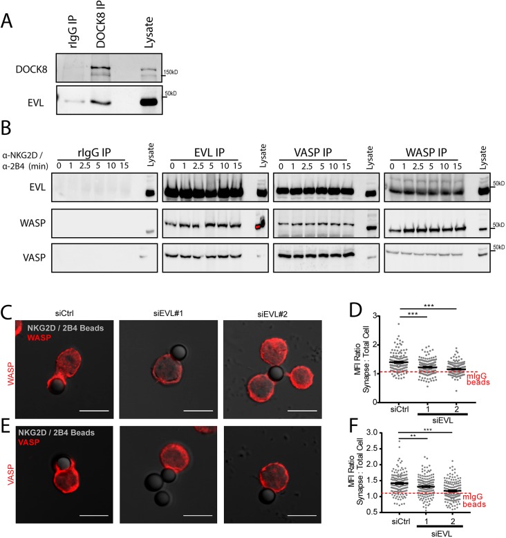
EVL is specifically required for the recruitment of WASP and VASP to the cytotoxic synapse. (A) NKL cells were lysed and immunoprecipitated (IP) with anti-DOCK8 antibody or rabbit IgG and immunoblotted for the indicated proteins. (B) NKL cells were stimulated over the indicated time course by ligation of NKG2D/2B4 receptors, and the indicated proteins were immunoprecipitated and immunoblotted. (C,E) NKL cells treated EVL siRNA (siEVL #1 or #2) or a control siRNA (siCtrl) were allowed to adhere to anti-NKG2D/2B4-coated latex beads for a total of 30 min, fixed and then imaged for the localization of (C) WASP and (E) VASP. Quantification of the MFI for (D) WASP and (F) VASP recruited to the cell–bead contact site from three independent experiments. Quantification is shown in relation to an average value for all three groups when conjugated to mIgG-coated latex beads, also in three independent experiments. Individual mIgG conjugate quantification can be seen in Fig. S4. Error bars indicate s.e.m. from the indicated mean **P<0.005; ***P<0.0005 (Student's t-test). Scale bars: 10 µm.

