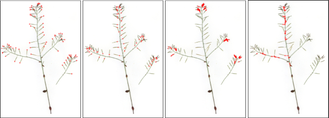
Figure 3.
Example annotated images (from left to right) for tip, base, body, and stem. Our annotation approach only requires sampling of pixels/points for the 4 main structural regions. Although most tips and bases have been annotated (see the left 2 panels), only a small portion of points for body or stems have been sampled and labelled (see the right 2 panels).

