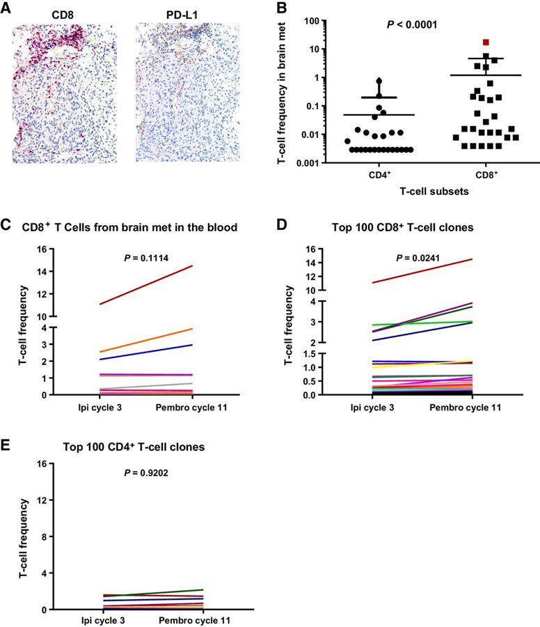
Figure 3.

Presence of immune infiltrate in brain metastases prior to immunotherapy and CD8+ T-cell expansion following pembrolizumab (Pembro). A, Immunohistochemical staining for CD8 and PD-L1 of a brain metastasis present prior to immunotherapy initiation. B-E, CDR3 sequencing of DNA derived from the brain metastasis FFPE, sorted CD3+CD4+ and CD3+CD8+ T cells from PBMCs, and bulk PBMCs. The bulk PBMCs were collected from ipilimumab (Ipi) cycle 3 and pembrolizumab cycle 11. B, The frequency of CD4+ and CD8+ T-cell subset in the brain metastasis at the same time point as in A. C, CD8+ T cell CDR3 sequences present in the brain metastasis were tracked in the blood at the two time points shown (ipilimumab cycle 3 and pembrolizumab cycle 11). D, The frequency of the top 100 CD8+ T-cell clones (in terms of frequency) was assessed at the same time points. E, Frequency of the top 100 CD4+ T cells clones across the two time points assessed.
