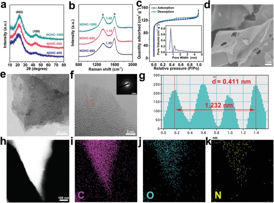
Figure 2.

Structural morphologies of NOHCs carbonized at different temperatures. a) XRD patterns and b) Raman spectra of NOHCs. c) N2 adsorption–desorption isotherms of NOHC‐800. The inset shows the pore size distribution of the adsorption branch obtained by the DFT method. d–f) FESEM, TEM, and the HRTEM images of NOHC‐800. The inset in f) is a SAED image of NOHC‐800. g) Line profile is acquired from the framed area in (f). h–k) High‐angle annular dark‐field scanning TEM image of NOHC‐800 and the corresponding elemental mappings for C, O, and N elements.
