
Figure 1.
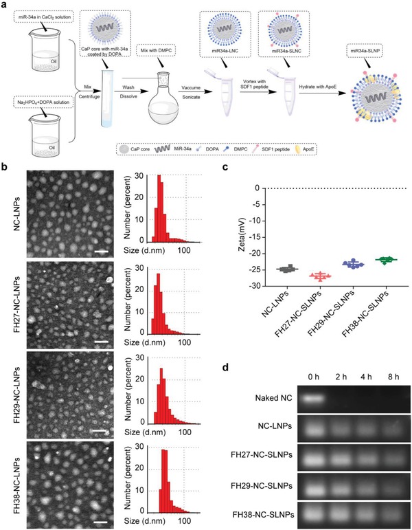
Preparation and characterization of NC‐SLNPs. a) The outline for the preparation of SLNPs nanoparticles loaded with microRNA. b) Morphology and particle size distribution of NC‐SLNPs (FH27/FH29/FH38) under a transmission microscope and dynamic light scattering. Scale bar, 50 nm. c) Particle zeta potential of NC‐SLNPs (FH27/FH29/FH38). d) Serum stability of miRNA loaded by LNPs and SLNPs at different incubation times.