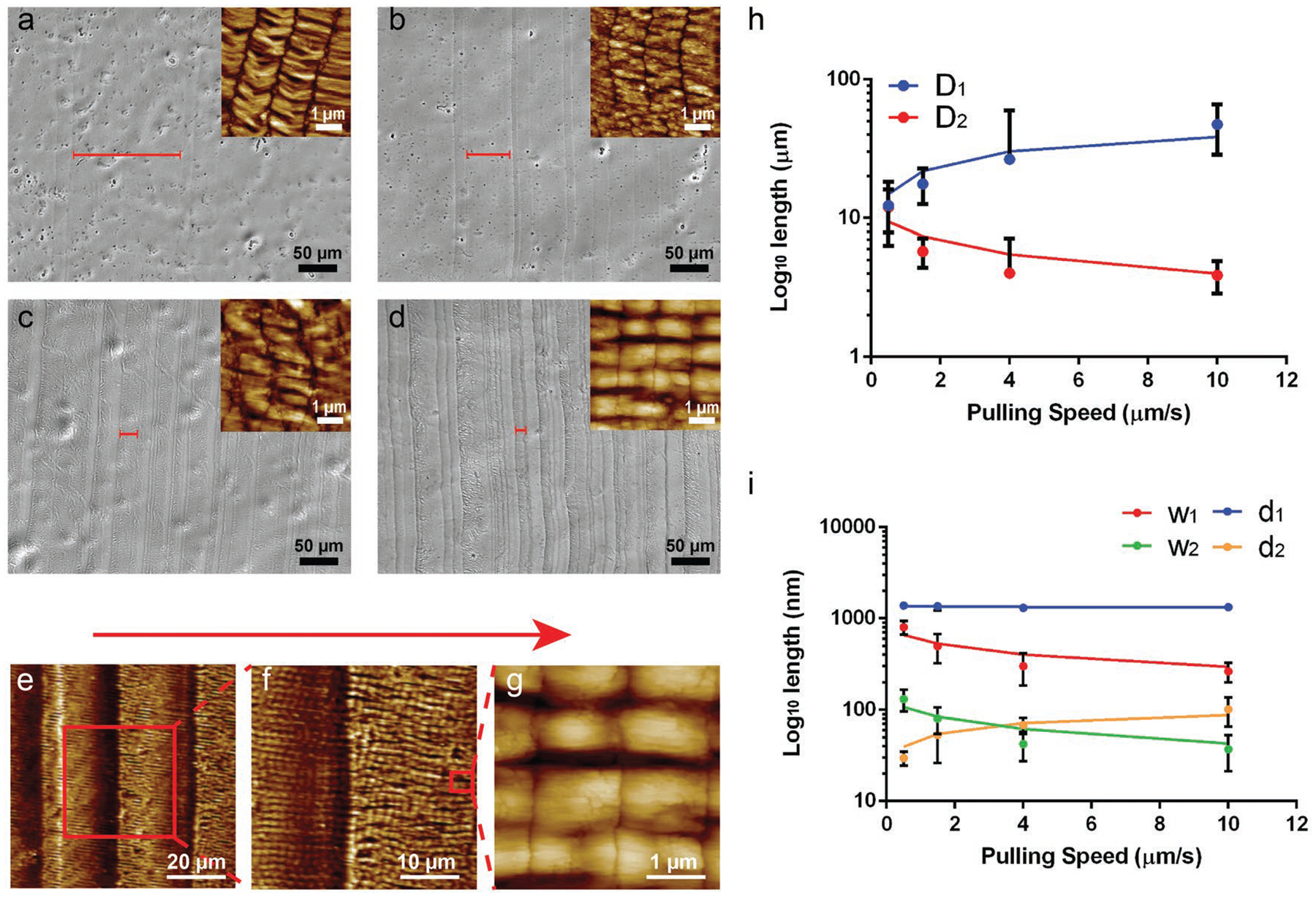
Figure 1.

Surface morphologies of ordered phage films assembled from a phage solution (1 × 10¹⁴ pfu mL−1) at different pulling speeds by the dip-pulling method. The surface morphologies were observed by a bright field optical microscopy imaging at a low magnification and by AFM imaging at a high magnification (shown as insets). a–d) The optical imaging of the film structures at different pulling speeds: a) 10 μm s−1; b) 4 μm s−1; c) 1.5 μm s−1; d) 0.5 μm s−1). The red bar on the images indicated the average width (D1) of the microridges at different speeds. As the speed decreased, the width (D1) of the microridges became smaller. e–g) AFM images of a NiM structure at different magnifications showing the microridges/microvalleys and nanoridges/nanogrooves (schematically shown in Scheme 1d). The red arrow denotes the pulling force direction. h,i), Relationship between the pulling speed and the six size parameters of the NiM (D1, D2, w1, w2, d1, d2). The length of d1 didn’t change with the speeds because it reflects the phage length, while the other five factors showed an increase (D1, d2) or a decrease (D2, w1, w2).
