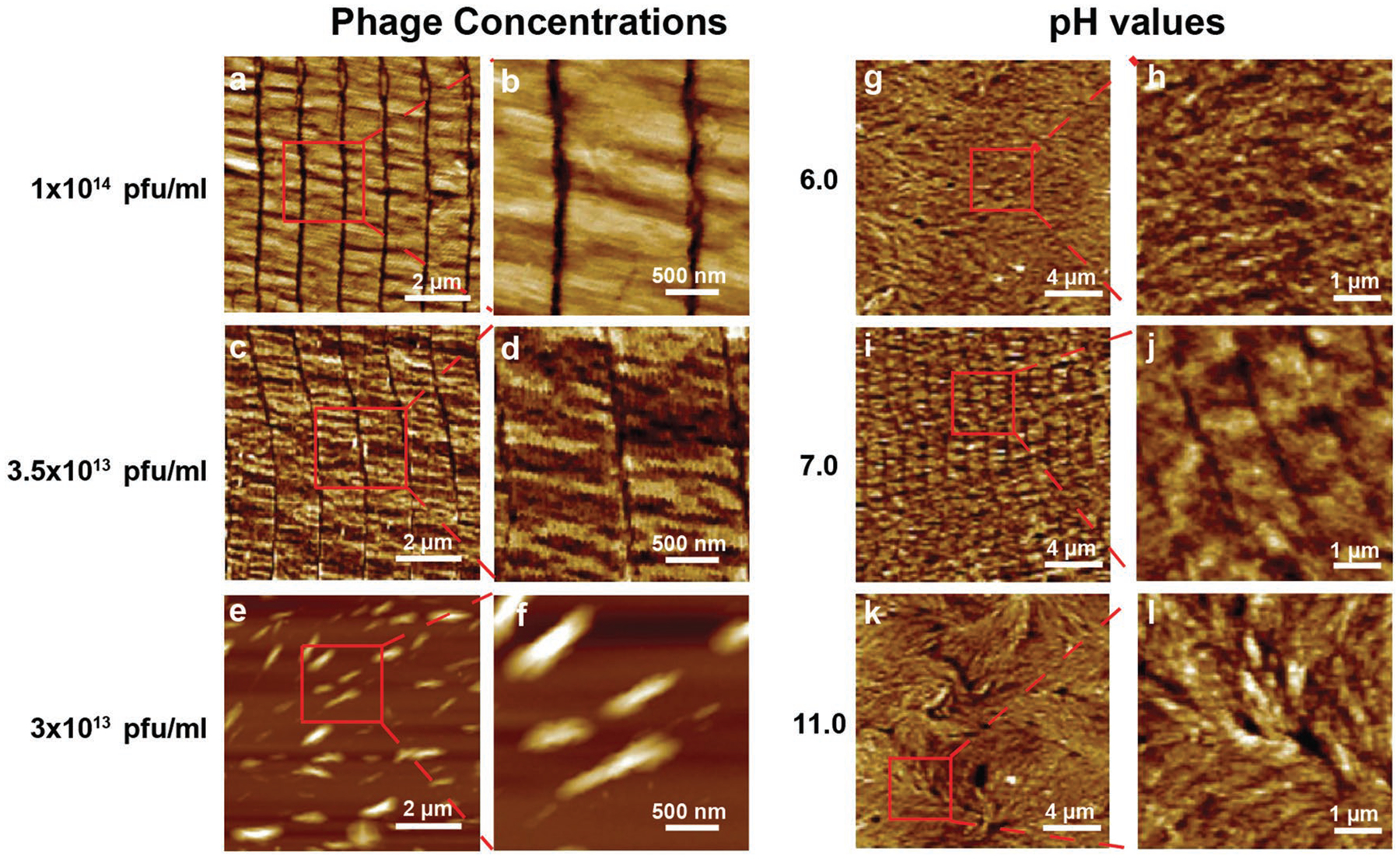
Figure 2.

Surface morphologies of wild-type phage films assembled form wild-type phages at different phage concentrations and pH values by the dip-pulling method. a–f) Phage concentration manipulates the phage film formation. The phages were assembled into similar ordered nanoridged patterns when their concentration was higher than 3.5 × 10¹³ pfu mL−1, below which such patterns were not formed. Pulling speed, 1.5 μm s−1. g–l) pH value of the phage solution manipulates the phage film formation. When the pH was between 6 and 11, the parallel-aligned nanoridged pattern could be seen under the AFM. When the pH was out of this range, such pattern was not formed. (Phage concentration, 5 × 10¹³ pfu mL−1; pulling speed, 1.5 μm s−1).
