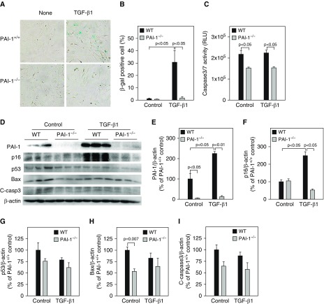
Figure 2.
Deletion of PAI-1 blocked TGF-β1–induced ATII cell senescence. ATII cells isolated from wild-type (WT) and PAI-1–knockout (PAI-1−/−) mice were treated with TGF-β1 as described in Figure 1. (A and B) SA-β-gal activity was revealed by X-gal staining. (C) Caspase 3/7 activity in the CM was measured with a kit from Promega. (D–I) Western blot analyses of the proteins of interest in ATII cells. (D) Representative Western blots. (E–I) Quantification of the band intensities, normalized by β-actin (n = 3–4).

