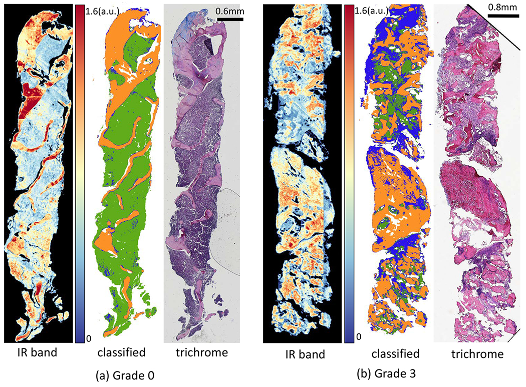
Figure 11.

Classification results of FT-IR imaged data for two biopsies with two collagen-deposition grades: (a) grade 0 and (b) grade 3. For each biopsy, IR image at band 1650 cm−1, pixel-level classified image with 16 optimal features for type I collagen (blue), trabecular bone (orange), and hematopoietic (green), and corresponding Mason’s trichrome stained adjacent tissue section.
