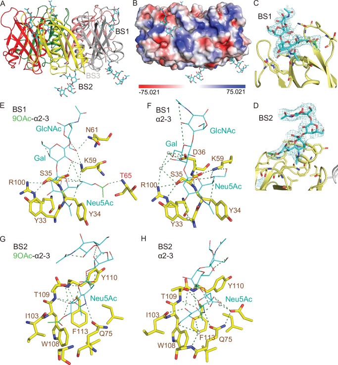
Fig 4. Co-crystal structure of S. Typhi PltB homopentamer bound to 9-O-acetylated α2–3 sialosides.
A, Crystal structure of PltB homopentamer in complex with Neu5,9Ac2α2-3Galβ1-4GlcNAc is shown as a ribbon cartoon with each protomer depicted in a different color. Cyan sticks, sugar carbon atoms. Blue sticks, nitrogen atoms. Red sticks, oxygen atoms. BS1-3, binding sites 1–3. B, Surface charge distribution of the PltB pentamer structure and the indicated glycan. C-D, Close-up view of the interface between the BS1 (C)/ BS2 (D) and the indicated glycans with their electron density maps. Green sticks, sugar carbon atoms of the 9-O-acetyl group. E-F, Close-up views of the BS1 complexed with Neu5,9Ac2α2-3Galβ1-4GlcNAc (E) or Neu5Acα2-3Galβ1-4GlcNAc (F). Dotted lines, H-bonds. Gray balls, water. G-H, Close-up views of the BS2 with 9-O-Ac α2–3 glycan (G) or unmodified α2–3 glycan (H).

