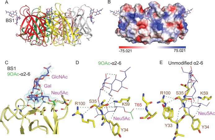
Fig 5. Co-crystal structure of S. Typhi PltB homopentamer bound to 9-O-acetylated α2–6 sialosides.
A, Crystal structure of PltB homopentamer in complex with Neu5,9Ac2α2-6Galβ1-4GlcNAc is shown as a ribbon cartoon with each protomer depicted in a different color. Purple sticks, sugar carbon atoms. Blue sticks, nitrogen atoms. Red sticks, oxygen atoms. BS1, binding site 1. B, Surface charge distribution of the PltB pentamer structure and the indicated glycan. C, Close-up view of the interface between the BS1 and the indicated glycan with the electron density map. Green sticks, sugar carbon atoms of the 9-O-acetyl group. D-E, Close-up views of the BS1 complexed with Neu5,9Ac2α2-6Galβ1-4GlcNAc (D) or Neu5Acα2-6Galβ1-4GlcNAc (E). Dotted lines, H-bonds.

