fig 3.

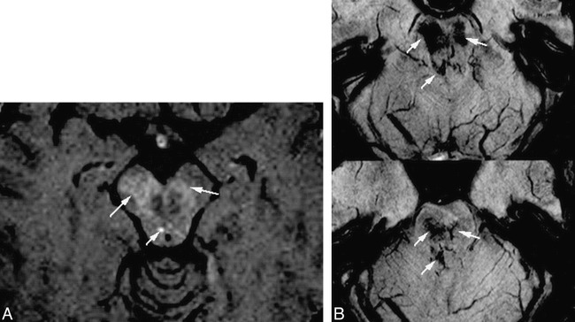
Comparison of HRBV and contrast-enhanced T1-weighted images.
A, Contrast-enhanced T1-weighted image (750/20 [TR/TE] with one acquisition) shows slight enhancement (arrows).
B, HRBV image shows abnormal vessels in the same regions compatible with telangiectasias. Some of the vessels have configurations of veins.
