
fig 6.
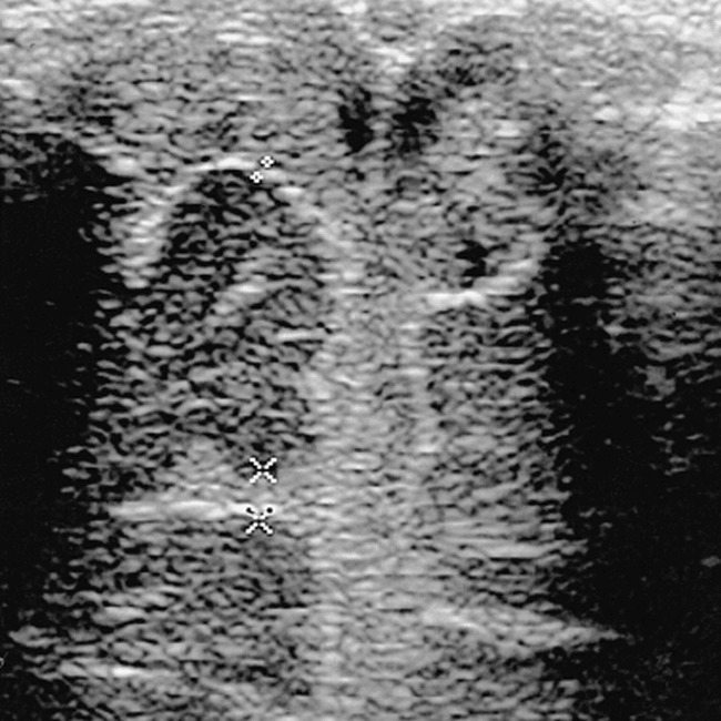
Coronal sonogram of a 3½-week-old infant with pneumococcal meningitis. The surface meninges are of normal thickness (0.8 mm). The subarachnoid space, the interhemispheric fissure, and the sulci are filled with echogenic material (pus and proteins by lumbar puncture). The frontal sulcus is enlarged (2.4 mm) and its undersurface is poorly defined