
fig 1.
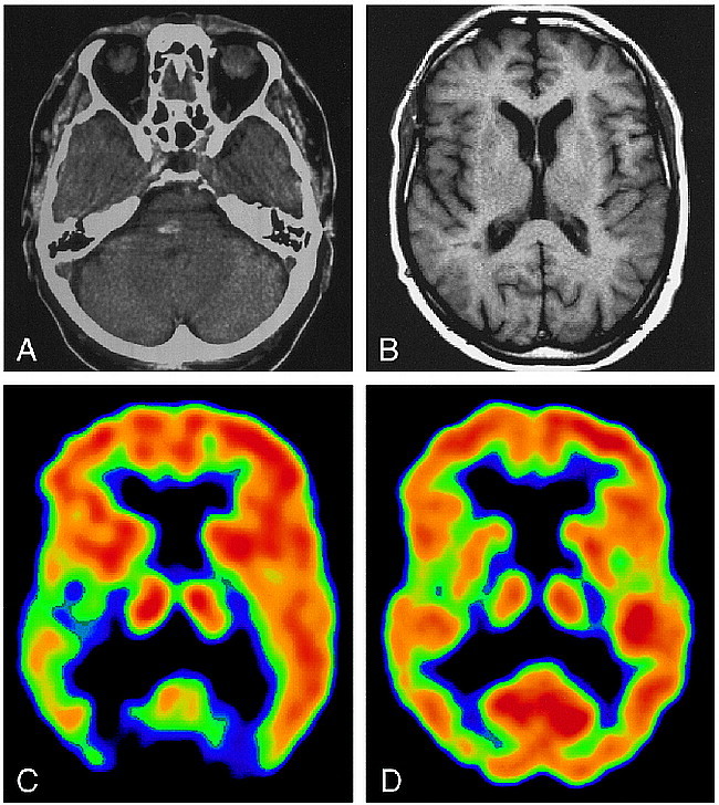
77-year-old patient with central poststroke pain.
A, CT scan on the day of the event shows a hyperdense lesion corresponding to a hemorrhagic vascular infarction.
B, MR image at the time of the PET study, at the same level as C and D.
C, IRF60 image shows a clear difference in opioid receptor binding between the hemispheres, with decreased activity on the right.
D, FDG image shows only slight differences in glucose utilization between left and right sides.