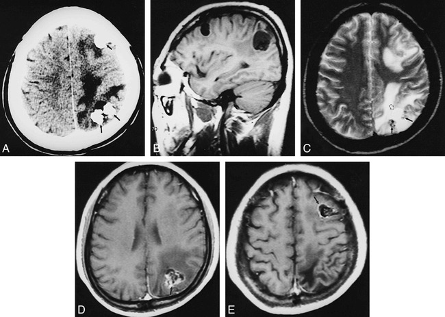
fig 2.

Case 2: 53-year-old man.
A, Noncontrast CT scan reveals multiple round calcifications (closed arrows) with eccentric cysts (open arrow) and moderate edema.
B, On T1-weighted image (420/14/2), the lesions show inhomogeneous hypo- and intermediate signal intensity in the left frontal and parietal lobes.
C, On T2-weighted image (2600/90/2), areas of heterogeneous signal intensity were noted in the left frontal and parietal lobes, caused by calcification (closed arrows), cyst (open arrow), and edema.
D and E, On contrast-enhanced T1-weighted image (420/14/2), the lesions show irregular enhancement (arrows).
