fig 1.

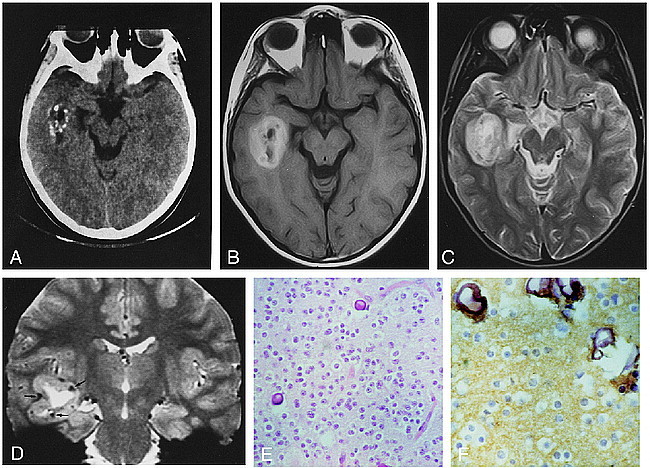
9-year-old girl with history of complex partial seizures.
A, On unenhanced CT scan, the lesion is centered in the right temporal lobe. Several calcified spots are visible in the peripheral part of the tumor. Central hypodense zones are consistent with cystic change. The noncalcified tumor parts are isodense with, and barely discernible from, adjacent brain. The apex of the right temporal horn is widened.
B, Axial SE T1-weighted image (600/16/2) shows the tumor mass centered in the white matter of the right temporal lobe. The lesion has a central hypointense core and a markedly hyperintense peripheral portion.
C, Axial SE T2-weighted image (2300/90/1) shows a markedly hyperintense peripheral portion. No perilesional edema is visible.
D, Coronal gradient-echo T2*-weighted image (1033/42/3; 20° flip angle) perfectly depicts the location of the mass in the right temporal lobe. The lesion has a large central cystic core and a peripheral solid part that is hyperintense to gray matter. Scattered, markedly hypointense foci in the peripheral part of the tumor (arrows) are consistent with calcified spots seen on the CT scan (see A).
E, Proliferation of round cells with clear cytoplasm associated with calcification (hematoxylin-eosin, ×25 magnification).
F, Intense staining with synaptophysin in the fibrillary background (×40 magnification).
