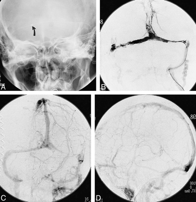
fig 1.

54-year-old woman with dural sinus thrombosis involving the superior sagittal sinus, both transverse sinuses, the right sigmoid sinus, and the right jugular bulb.
A, Plain frontal radiograph shows the AngioJet catheter (navigated through the left internal jugular vein) positioned in the mid right transverse sinus. Arrow indicates catheter tip.
B, Frontal venogram, obtained through the AngioJet catheter after rheolytic thrombectomy, shows partial interval sinus thrombolysis.
C and D, Frontal (C) and lateral (D) venous phases from a left internal carotid angiogram, performed after completion of rheolytic thrombectomy and urokinase thrombolysis, show restored patency of the major dural venous sinuses.
